


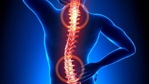
Excessive tension of the back muscles causes a lot of discomfort and pain. Osteochondrosis, which causes a violation of the structure of the vertebrae and intervertebral discs, leads to serious pinching of the nerve endings. Often, the pathology is accompanied by a deterioration in blood circulation, which provokes disruptions in the nutrition of the brain and internal organs.
Osteochondrosis - what is it?

Osteochondrosis is a recurrent type of disease that occurs in a chronic form and is accompanied by destruction of the vertebrae with intervertebral discs. Their tissues are disturbed, which provokes a decrease in the degree of their elasticity with a subsequent change in shape. There is a gradual decrease in the intervertebral space. This causes a loss of stability of the spinal column in areas of pathology development.
The processes of pathological tissue destruction occur against the background of pinched nerve endings, which are directed from the area where the spinal cord is located. As a result, the back muscles are in constant tension. In such a situation, patients complain of pain in the back and other symptoms.
Based on the peculiarities of the localization of the structures of the spine, which were covered by degenerative changes, the cervical, thoracic and lumbosacral types of the pathological process are distinguished. The main symptom of the development of osteochondrosis is pain, the intensity and severity of which usually increases during physical exertion.
There is also stiffness in movement. In addition, the clinical picture is characterized by the presence of signs of the vertebral type - headaches, changes in blood pressure, deterioration in visual function, hearing, and so on.
Development Mechanism
The development of osteochondrosis is associated with the fact that the nucleus pulposus begins to lose its hydrophilic qualities. This semi-liquid structure contains connective tissue fibers and chondroitin, a gelatinous substance. In the process of development of the human body and its growth, the processes of reduction of the vascular bed in the intervertebral discs are actively proceeding. Nutrients are supplied in a diffuse manner, which manifests itself in the spontaneous stabilization of the concentration. This feature becomes the reason for difficulties in the complete restoration of cartilage that has suffered damage or excessive pressure on the spine.
Pathological abnormalities become more striking due to violations in the hormonal background and human nutrition. The cartilage tissue begins to lack nutrients that are required for its normal development. Therefore, disorders appear in the form:
- decrease in strength and elasticity;
- changes to consistency parameters and configuration properties.
Against the background of flattening of the intervertebral discs, the formation of radial cracks in the fibrous rings occurs. As a result, the intervertebral distance is reduced, and the facet joints begin to shift. Over time, pathological changes cover connective tissue types related to the fibrous rings and ligaments.
As tissues break down by the immune system, increased amounts of immunoglobulins are produced. This provokes the development of the process of aseptic inflammation, edema is formed in the area where the facet joints are located. They also spread to the adjacent soft tissues.
Due to the stretching of the joint capsules, the intervertebral discs lose their ability to fix the vertebrae. Such instability of the position of the structure of the spine increases the risks of pinching of the nerve roots or squeezing of blood vessels. This feature is typical, for example, for cervical osteochondrosis, which is accompanied by intense verbal symptoms.
Causes of the disease
The condition of the intervertebral discs can worsen with a decreased tone of the skeletal muscles in the spine. Due to the irrational and asymmetric work of the muscles, destruction of cartilaginous tissues may occur with the prolonged preservation of the non-physiological position of the body. This violation is the result of wearing heavy bags on the same shoulder, using soft mattresses and high pillows.
The process of destruction of intervertebral discs is accelerated due to the action of a number of negative factors of external and internal nature. These include:
- disorders of the endocrine mechanism and metabolic disorders;
- pathologies of an infectious nature, including in a chronic form;
- injuries of the spinal column in the form of compression fractures, bruises;
- regular and prolonged hypothermia of the body;
- diseases of systemic and degenerative-dystrophic type - gouty, psoriatic, rheumatoid arthritis, osteoporosis, osteoarthritis;
- smoking and alcohol abuse, which disrupts the state of the vascular system, impairs blood circulation and provokes a lack of nutrients in the cartilage;
- insufficient physical development, problems with posture, flat feet - these defects increase the load on the spinal column, since the amortization will be insufficient;
- obesity;
- genetic predisposition;
- exposure to regular stress.
Symptoms
The main clinical sign of osteochondrosis of any localization (cervical, thoracic or lumbosacral) is pain syndrome. With a relapse, the pain is penetrating, radiating to the nearby areas of the body. Even with a slight movement, it intensifies. This forces the patient to put the torso in a forced position in order to minimize discomfort and soreness:
- with cervical osteochondrosis, it will be preferable to turn not one head, but the whole body;
- when the chest form of the disease is present, it is difficult for the patient to take a deep breath, and therefore, in order to exclude acute pain in the chest, he tries to minimize the depth and frequency of breathing;
- in patients with a lumbar type of disease, difficulties arise when they sit down, take an upright position, move, as the nerve is pinched in the spinal location.
Typically, patients complain of the presence of dull, persistent pain and a feeling of stiffness in movements in the morning after waking up. In this case, differential diagnosis will be required to help eliminate the risks of developing myositis caused by inflammation of the skeletal spinal muscles or osteoarthritis.
Aching and pressing pains occur due to compensatory tension in muscle tissues. This condition is necessary to stabilize the spinal motion area. Constant mild or moderate pain can appear with a significant stretching of the intervertebral disc, and result from aseptic inflammatory changes.
Osteochondrosis of a separate localization is characterized by special symptoms:
- With cervical osteochondrosis, pain is felt in the cervical zone, in the upper limbs. Pains in the head and numbness of the fingers are observed. If the disease manifests itself in a serious form, then pinching of the vertebral artery can occur. In this case, the patient begins to complain of a significant deterioration in health.
- Thoracic localization is manifested by acute and aching pain in the back, visceral pain syndrome is present in the cardiac region, right hypochondrium and abdomen. Patients complain of numbness, paresthesia of the skin, shortness of breath, crunching in the vertebrae.
- Patients with lumbar osteochondrosis complain of pain in the back and lower extremities with increased intensity when moving. Often, disorders in the functioning of the genitourinary system, problems with male potency, and dysfunctional ovarian disorder are diagnosed. During remission, pain may decrease. However, the impact of a provoking factor leads to its renewal.
- When mixed osteochondrosis manifests itself, the symptomatology can manifest itself in several zones at the same time. This condition is characterized by a more severe course of the disease.
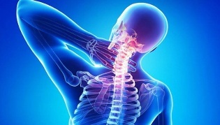
It should be remembered that the displacement of the vertebrae and the formation of osteophytes cause compression of the vertebral artery. It nourishes the brain, providing its cells with an oxygen component. When squeezed, food is limited, and therefore the patient has problems with coordination, headaches, tinnitus, and arterial hypertension.
Consequences if untreated
The reason for the complicated course of osteochondrosis is the relatively rapid formation of hernias in the intervertebral discs. Their appearance is associated with the displacement of the vertebral structure in the posterior direction. This provokes a rupture of the posterior ligament of the longitudinal type, which results in instability of the position of the disc, protrusion of its individual sections into the region of the spinal canal. Rupture of a hernia occurs when a disc with a nucleus pulposus penetrates into the canal area.
With the manifestation of pathological abnormalities in the vertebral structures, the back of the brain begins to squeeze, the patient develops discogenic myelopathy. The symptoms of this condition are associated with numbness and weakness in certain muscle groups of the upper and lower extremities. Paresis, muscle atrophy, and tendon reflexes are manifested. In some cases, there are problems with emptying the bladder, with the intestines.
Herniated discs are dangerous by squeezing the arteries that supply the spinal cord. The result of this pathology is the formation of ischemic zones, where nerve cells have undergone damage and death. The manifestation of the neurological effect is expressed in malfunctions of motor function, a drop in the degree of tactility, and a disorder of trophism.
Disease Diagnostics
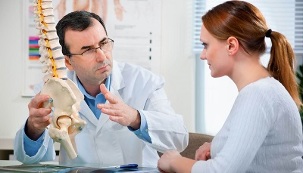
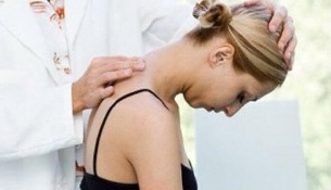
The initial diagnosis is made on the basis of the patient's complaints and the existing symptoms. The specialist studies the condition of the spinal column in different positions, suggesting that the patient be at rest or in motion. At the next stage, the patient is directed to laboratory diagnostics, which will help clarify the diagnosis or refute it.
The methods of the applied research include:
- Radiography- provides a complete examination of the spinal column with an assessment of the condition of the vertebrae, existing disorders in the form of growths, curvatures. The specialist will be able to determine the intervals of the intervertebral type, the state of the holes. To accurately identify osteochondrosis, localized in the chest or cervical area, a two-stage x-ray examination is performed. In the first stage, the patient lies on his side, and in the second, directly on his back.
- The method of tomography by means of MRI or CTgives highly informative data, which helps to study the vertebrae in detail without interference in the form of organs covering them. The picture shows the nerves and the vascular system. MRI helps to identify the signs of many diseases of the spinal column and the location of the damage. With CT, hernias are visualized, possible deviations in the structure of the spine are determined.
- Laboratory examinationto assess the condition of the blood and its main parameters. Allows you to clarify the diagnosis and determine the possibility of developing concomitant diseases.
In many cases, as a result of examinations, doctors diagnose the presence of some background diseases, potentially dangerous for their complications. We are talking, for example, about hernias, protrusion, radiculitis. Correct diagnosis of problems helps to effectively treat osteochondrosis. At the same time, the disease itself in the early stages of development is disguised as the symptoms of other diseases.
Therapeutic Process
Osteochondrosis is treated conservatively or with surgery. The choice depends on the severity of the condition, its neglect, the level of tissue deterioration, and the causes.
It is important to remember that it is not possible to completely cure osteochondrosis, since there are no medications to help completely restore discs and vertebrae. The therapeutic effect is focused on inhibiting the destruction process and increasing the duration and stability of remission.
For symptomatic therapy, chondroprotectors are used, which are based on chondroitin sulfate or glucosamine.
The effectiveness of the therapeutic process with the use of chondroprotectors has been clinically confirmed on the basis of long-term tests. If you take these funds for a long time from 3 months, then there is a partial restoration of cartilage and other elements of the connecting type - the ligamentous-tendon apparatus, bursa.
The accumulation of glucosamine and chondroitin in the intervertebral disc area leads to the manifestation of analgesic, anti-edematous and anti-inflammatory effects. Therefore, there is a real opportunity to optimize the dosage of NSAIDs, drugs of the glucocorticosteroid group, muscle relaxants. You can count on a decrease in the drug load on the patient.
The effectiveness of chondroprotectors is determined by the regularity of their intake. Otherwise, there will be no result. Ineffectiveness is also recorded in the treatment of grade 3 osteochondrosis, accompanied by significant destruction of cartilage.
The following groups of drugs can be used to relieve pain:
- Non-steroidal anti-inflammatory drugshelp to eliminate inflammatory disorders in soft tissues that are caused by vertebral displacement. NSAIDs are effective in reducing pain, swelling, and stiffness.
- Means of the glucocorticosteroid group- usually blockades are used in conjunction with an anesthetic. They are able to relieve pain, restore the immune mechanism, and provide an anti-exudative effect.
- Muscle relaxants.They are effective in combating muscle spasms due to nerve entrapment. They help to relax the muscles of the skeleton and block reflexes of the polysynaptic spinal type with an antispasmodic effect.
- External remedies with warming effect.Irritation of subcutaneous tissue receptors with activation of blood flow is provided by special gels and ointments. These drugs have analgesic and anti-edematous effects.
It is possible to eliminate the symptoms of the vertebrogenic type, manifested as a result of the localization of pathology in the cervical or thoracic zone, with the help of medical devices to activate blood flow. Nootropics and drugs to improve microcirculation are also prescribed. In some cases, you may need to take antidepressants, as well as pharmaceuticals with anticonvulsants.
During the treatment of osteochondrosis, physical therapy is also used. The procedures of UHF therapy, magnetotherapy, laser therapy, reflexology, massage, exercise therapy, hirudotherapy, as well as swimming and yoga can be prescribed. If conservative treatment is ineffective, the operation is performed using microdiscectomy, puncture disc valorization, laser reconstruction or implant replacement.